3d lung ct deep learinig
Addressing challenges in other common imaging modalities gruezemacher et al developed a three-dimensional 3d adaptation of deep neural nets dnns for lung nodule detection from computed. 1 A deep active learning-based nodule detection framework is proposed to achieve comparable performance with less data and annotations as shown in Fig.
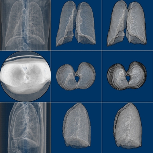
3d Lung Segmentation University Of Dayton Ohio
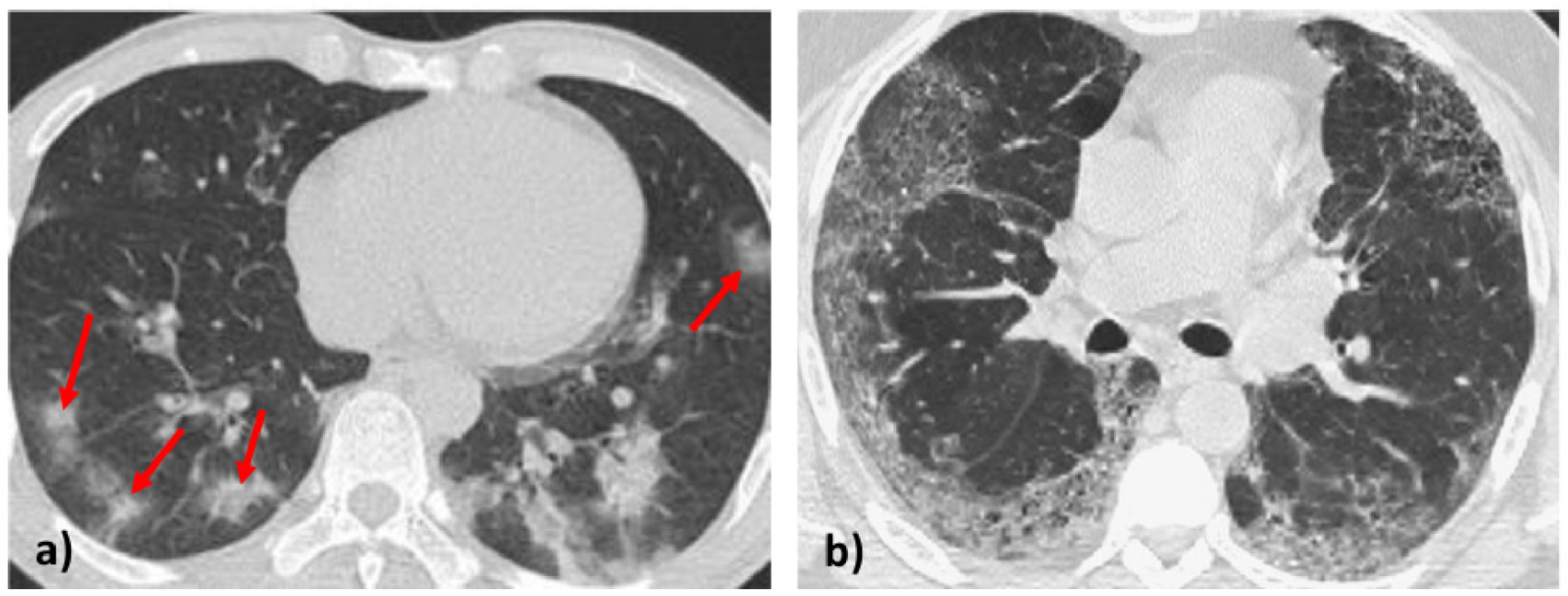
Pneumonia is the inflammation of the alveoli inside the lungs 3.
. Towards radiologist-level cancer risk assessment in CT lung screening using deep learning. The purpose of this study is to compare the detection performance of the 3-dimensional convolutional neural network 3D CNN-based computer-aided detection CAD models with radiologists of different levels of experience in detecting pulmonary nodules on thin-section computed tomography CT. Harbert College of Business Auburn University Ashish Gupta PhD.
Using 5-fold cross validation a mean Dice coefficient of 0988 0012 and Average Surface Distance of 0562 049 mm was achieved by the proposed 2D CNN on lung segmentation. Deep-Learning framework for COVID-19 chect CT analysis Image by author 1. The arrows of different colors.
Lung Nodule Detection With Deep Learning in 3D Thoracic MR Images Abstract. Slice based solution CT scan include a series of slices for those who are not familiar with CT read short explanation below. A VGG19-based encoder path Shallow U-DL upper and b deep DenseNet-based encoder path Deep U-DL bottom.
In this work we investigate whether augmenting a dataset with. By Stojan Trajanovski et al. Antibiotics and antiviral drugs can treat bacterial and viral pneumonia.
Sharp High Contrast Resolution. The patient will show symptoms such as shortness of breath cough fever chest pains chills or fatigue. Recently it has been demonstrated that screening those at high.
AiCE is an innovative Deep Learning Reconstruction technology thats been trained to reduce noise and boost signal to deliver sharp clear and distinct images at speed. The size of each feature map is shown at the lower-left edge of the box. Clear Low Contrast Detectability.
Researchers validated the algorithm using an additional 2085 NLST. This challenge is especially acute within the medical image domain particularly when pathologies are involved due to two factors. 3D DenseNet on lobar segmentation achieved Dice score of 0959 0087 and Average surface distance of 0873 061 mm.
The architectures of our two asymmetric U-shaped deep learning backbone networks for lung nodule segmentation in CT images. We build a small yet definite CT dataset 171 patients called SCH-LND focusing on solid lung nodules 90 benign90 malignant cases. 3D DEEP LEARNING FOR DETECTING PULMONARY NODULES IN CT SCANS 3D DEEP LEARNING FOR DETECTING PULMONARY NODULES IN CT SCANS Ross Gruetzemacher Doctoral Student Department of Systems Technology Raymond J.
Share Lung cancer is the leading cause of cancer mortality in the US responsible for more deaths than breast prostate colon and pancreas cancer combined. 1 limited number of cases and 2 large variations in location scale and appearance. As a result our approach allows us to determine CT images in the middle lung region of the 3D volume.
Data availability plays a critical role for the performance of deep learning systems. The inflammation will build up fluid and pus that subsequently causes breathing difficulties. Early detection of lung cancer is crucial in reducing mortality.
Keywords CT Lung segmentation Lobar. AiCE is available on. The proposed deep learning method First the method selects middle axial lung slices Section 31 and then each of these middle axial slices goes through ResNet-50 convolutional neural network CNN model.
To address the above challenges this paper makes the following contributions. Using a large dataset from the National Lung Screening Trial NLST Yan and his team used data from more than 30000 low-dose CT images to develop train and validate a deep learning algorithm capable of filtering out unwanted artifacts and noise and extracting features needed for diagnosis. Accordingly in this paper we present a framework for identifying pulmonary nodules in lung CT images and a convolutional neural network CNN approach to automatically extract the.
Magnetic resonance imaging MRI may be a viable imaging technique for lung cancer detection. Numerous lung nodule detection methods have been studied for computed tomography CT images. Machine learning and image processing techniques generally embedded in computer-aided diagnosis CAD systems might help radiologists locate and assess the risk of these nodules.
The purpose of this study is to compare the detection performance of the 3-dimensional convolutional neural network 3D CNN-based computer-aided detection CAD models with radiologists of different levels of experience in detecting pulmonary nodules on thin-section computed tomography CT. LUNG NODULE DETECTION IN CT USING 3D CONVOLUTIONAL NEURAL NETWORKS Xiaojie Huang Junjie Shan and Vivek Vaidya GE Global Research Niskayuna NY ABSTRACT We propose a new computer-aided detection system that uses 3D convolutional neural networks CNN for detect-ing lung nodules in low dose computed tomography. Under the supervision of SCH-LND dataset many hidden drawbacks of unsure data 484 solid nodules selected from LIDC-IDRI dataset served for malignancy prediction are objectively revealed.
The unlabeled CT scans are assessed by a trained model with predicted uncertainties. Since we had a very limited number of COVID-19 patients scans we decided to use 2D slices instead of 3D volume of each scan.
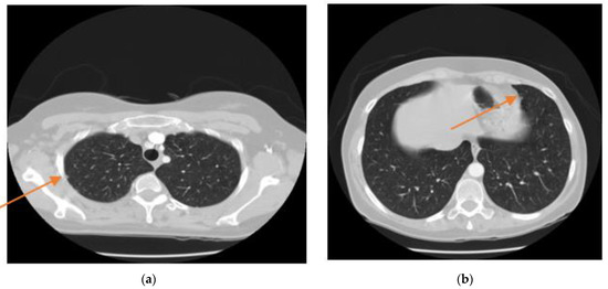
Sensors Free Full Text Resbcdu Net A Deep Learning Framework For Lung Ct Image Segmentation Html
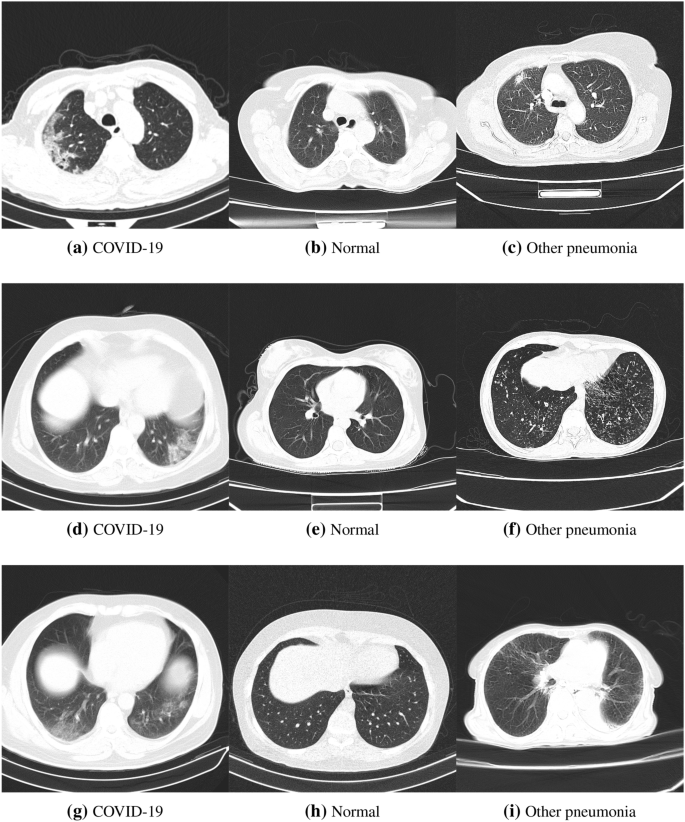
Proposing A Novel Deep Network For Detecting Covid 19 Based On Chest Images Scientific Reports
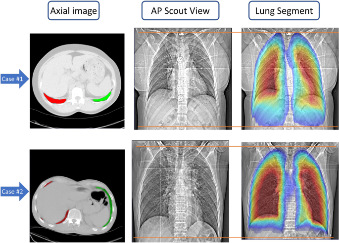
Deep Learning Based Fully Automated Z Axis Coverage Range Definition From Scout Scans To Eliminate Overscanning In Chest Ct Imaging Insights Into Imaging Full Text
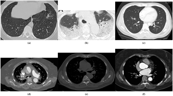
Proposing A Novel Deep Network For Detecting Covid 19 Based On Chest Images Scientific Reports

Detection And Analysis Of Covid 19 In Medical Images Using Deep Learning Techniques Scientific Reports
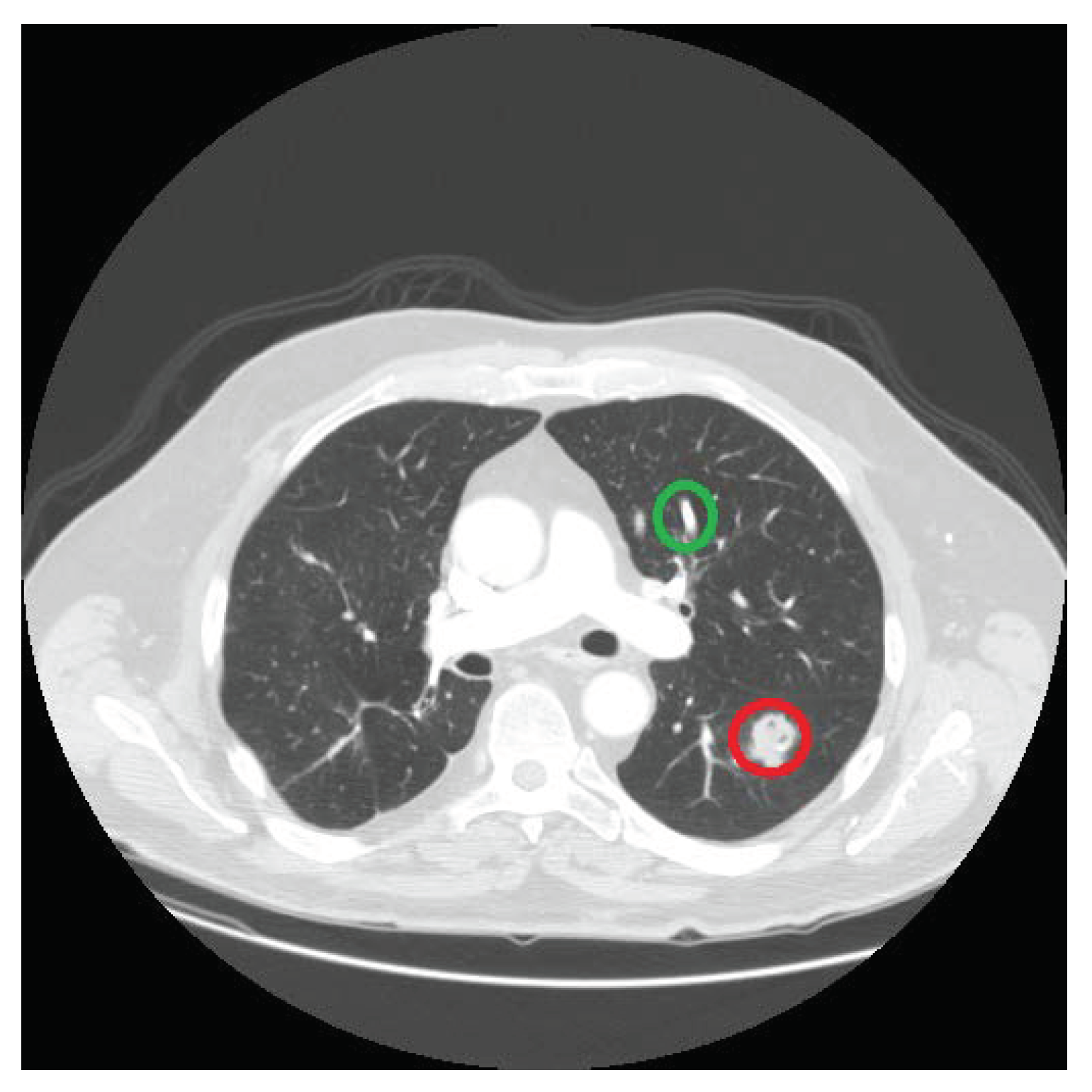
J Imaging Free Full Text Lung Nodule Detection In Ct Images Using Statistical And Shape Based Features Html
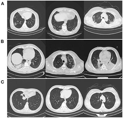
Frontiers Covid Net Ct 2 Enhanced Deep Neural Networks For Detection Of Covid 19 From Chest Ct Images Through Bigger More Diverse Learning

Lung Cancer Detection From Ct Image Using Improved Profuse Clustering And Deep Learning Instantaneously Trained Neural Networks Sciencedirect
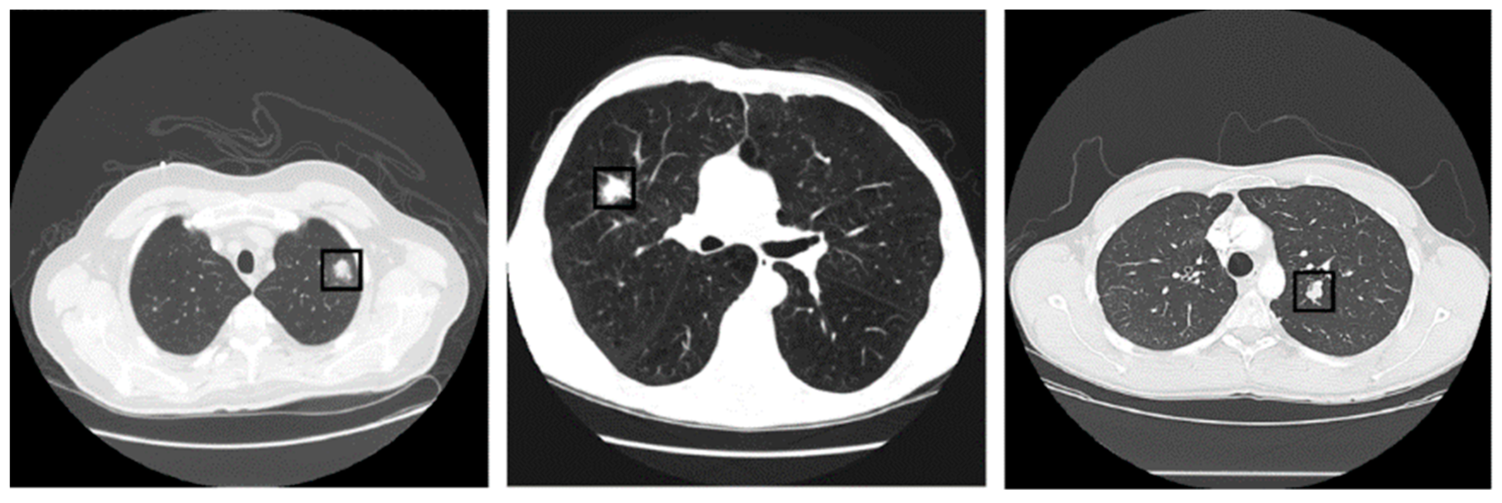
Sensors Free Full Text Automated Lung Nodule Detection And Classification Using Deep Learning Combined With Multiple Strategies Html

Multi Task Deep Learning Based Ct Imaging Analysis For Covid 19 Classification And Segmentation Medrxiv
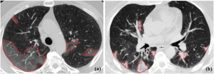
Covid Rate An Automated Framework For Segmentation Of Covid 19 Lesions From Chest Ct Images Scientific Reports
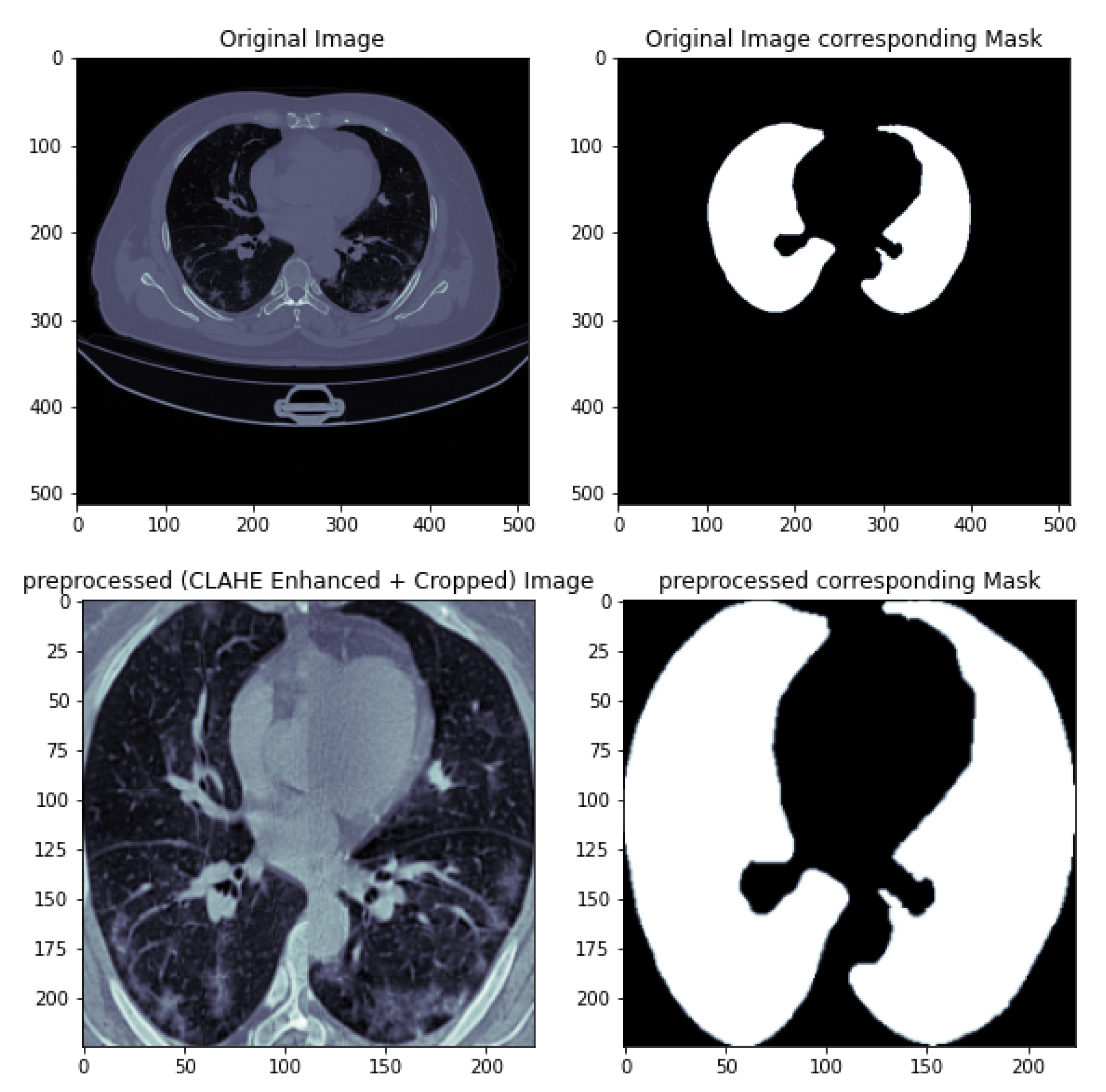
Applied Sciences Free Full Text A Deep Learning Based Diagnosis System For Covid 19 Detection And Pneumonia Screening Using Ct Imaging Html

Deep Learning Analysis Provides Accurate Covid 19 Diagnosis On Chest Computed Tomography European Journal Of Radiology

Applied Sciences Free Full Text Deep Learning For Covid 19 Diagnosis From Ct Images Html
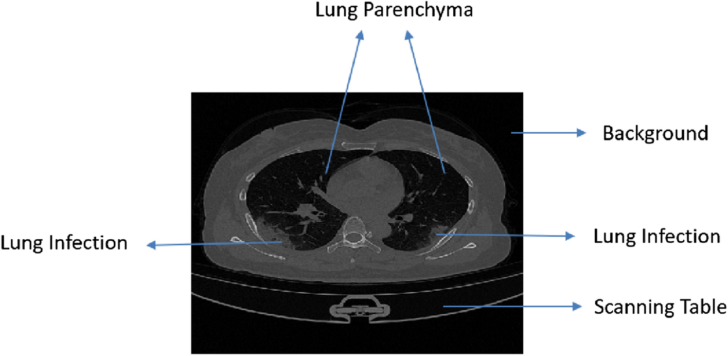
Cascaded 3d Unet Architecture For Segmenting The Covid 19 Infection From Lung Ct Volume Scientific Reports
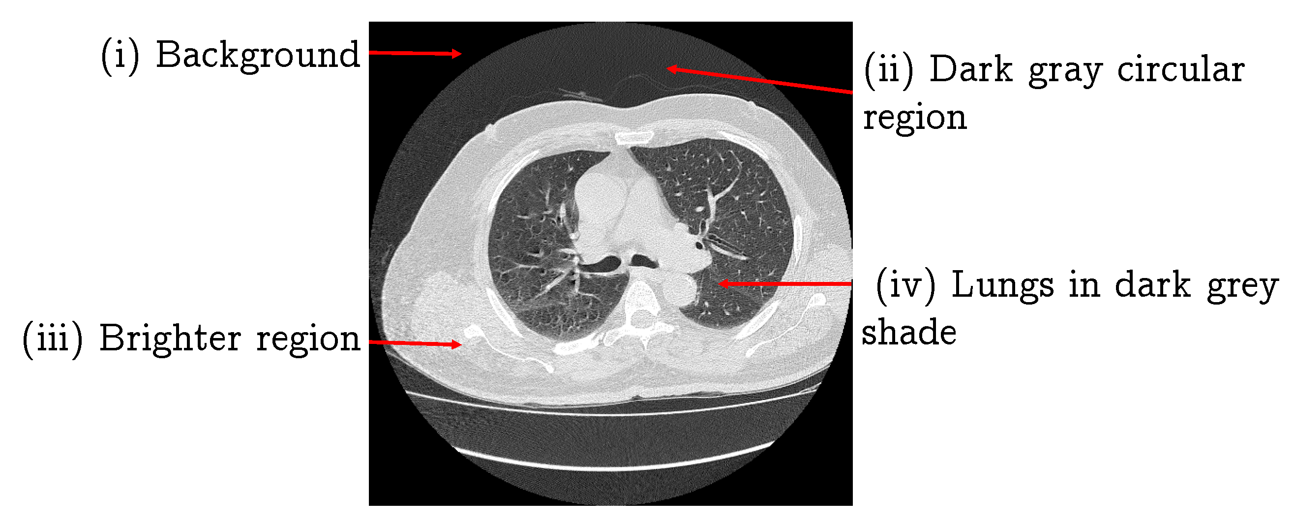
J Imaging Free Full Text Lung Nodule Detection In Ct Images Using Statistical And Shape Based Features Html
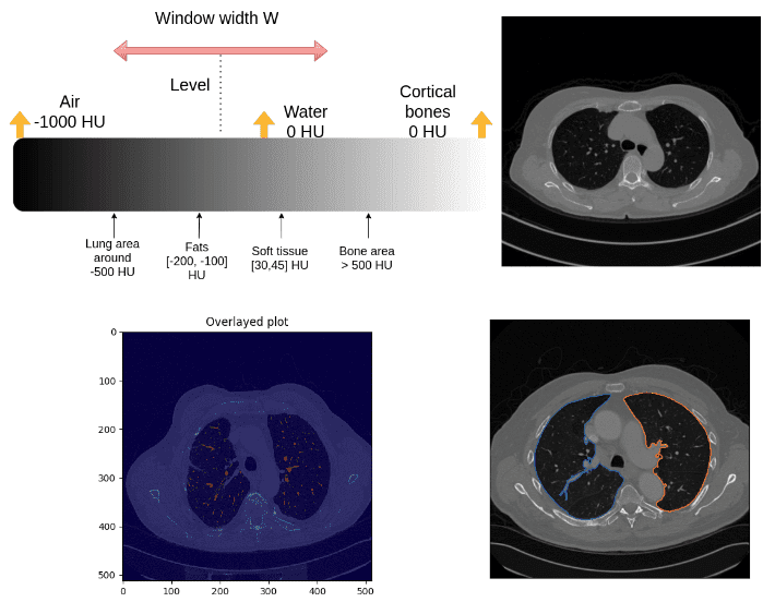
Introduction To Medical Image Processing With Python Ct Lung And Vessel Segmentation Without Labels Ai Summer
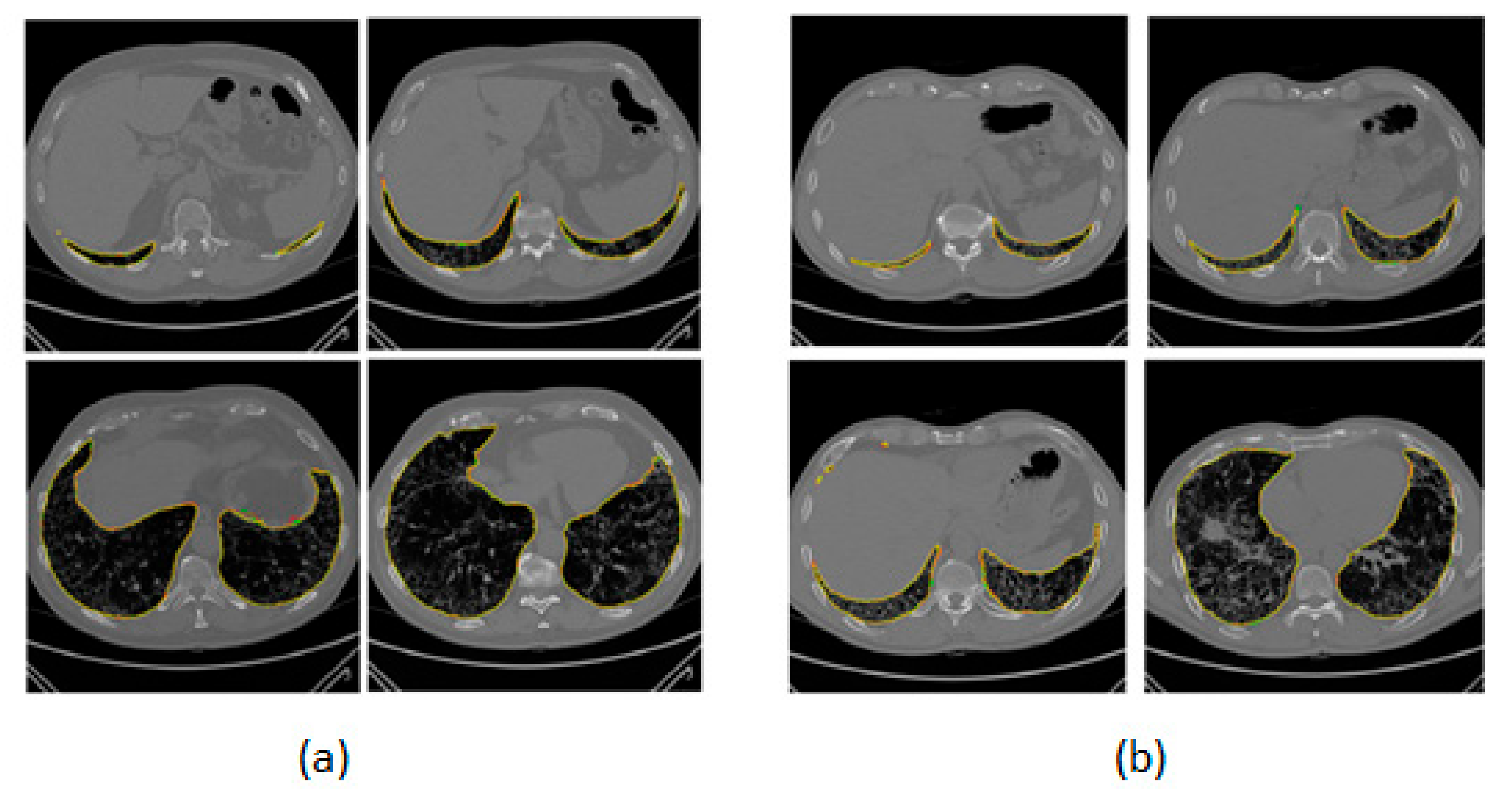
J Imaging Free Full Text Lung Segmentation On High Resolution Computerized Tomography Images Using Deep Learning A Preliminary Step For Radiomics Studies Html